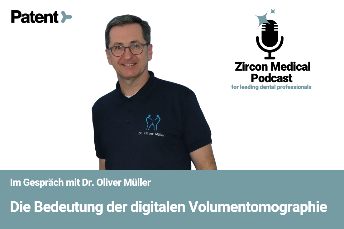
Dr. Oliver Müller
Lecturer in the Field of Digital Volume Tomography
Graduated in 1994 from the Johannes-Gutenberg University in Mainz
Working in the joint practice of Dr. Mirjam Frech and Dr. Fabian Schach since April 2022
Founded the company GO Dentale Fortbildung und Beratung
Lecturer in the field of three-dimensional imaging in X-rays — DVT (Digital Volume Tomography)
YouTube: https://www.youtube.com/channel/UCsBJF610oCdEcuSb2shamyA/featured
LinkedIn: https://www.linkedin.com/in/oliver-m%C3%BCller-288023151/
DVT Courses: http://www.dvt-mueller.de
Nelkenstraße 1, 78532 Tuttlingen, Germany
In Conversation with Dr. Oliver Müller
Technological innovations over the past decade or two have dramatically transformed the diagnostic and therapeutic possibilities within dentistry. Sometimes, technological changes outpace how quickly dentists can adapt to the changes — three-dimensional imaging with DVT (digital volume tomography) is a prime example of one such technological innovation.
Our team at Zircon Medical recently hosted Dr. Oliver Müller, a dentist from Tuttlingen who specializes in 3D imaging, on our podcast series to discuss the importance of integrating digital volume tomography in dental clinics.
Introducing Dr. Oliver Müller, a lecturer in the field of digital volume tomography
Dr. Oliver Müller is a dentist from Tuttlingen in Germany. He previously ran a joint practice with Dr. Mirjam Frech for a total of 26 years. But as of April 2022, he took a step back within the practice to focus more on being a lecturer within the field of digital volume tomography (DVT), a role that he’s been fulfilling since 2009. The practice is now run by Dr. MirjamFrech and Dr. Fabian Schach, and Dr. Müller is giving more time to being a lecturer.
Physical and online training courses for DVT
Dr. Müller also offers online training for DVT. In Switzerland, dentists must have a DVT license to use the technology, but because of the pandemic-induced travel limitations, dentists couldn’t access physical training. That paved the way for the mainstreaming of online training courses — Dr. Müller offered online courses to meet that demand. But now that the pandemic is drawing to an end, Dr. Müller isn’t sure if the authorization to offer DVT licenses online will continue.
Dr. Müller says purely theoretical training can be fairly dry, and certain aspects of dental radiography are hard to teach online. But software training can be handled online — he has also made numerous tutorials on his YouTube channel DVT-Müller. Over the past year and a half, Dr. Müller has noticed that younger colleagues generally prefer online training to avoid travel costs and efforts, but older colleagues generally prefer face-to-face events.
Dr. Müller limits online training events to 15 participants because that allows him to engage with each individual. He can see all the participants on the screen, and he tries to respond to each person individually. Within this framework, he says patients ask all kinds of questions, and he can offer individualized support to everyone, which is greatly appreciated.
What to expect from Dr. Müller’s DVT course
Dr. Müller’s online training course lasts for two days. The first day focuses primarily on theoretical knowledge about radiation protection and how the DVT device works. The class explores the classes of equipment available, how different devices work, and the indications for usage. Dental radiography has numerous indications that go beyond dental surgery and implantology, and Dr. Müller explores those indications during the training event.
The DVT course also includes software training and homework for the participants. The participants must assess 25 data sets of DVT according to a deadline formulated by the Federal Office for Radiation Protection. The participants have three months to complete the homework, following which they appear for the second course, which deals with further theoretical knowledge. On the second day, Dr. Müller discusses the possibilities of misdiagnosis and other dangers.
Dr. Müller also demonstrates the software to his participants. He says the specific software they use is of secondary importance. “If you are quite fit in one of the programs, you will also succeed relatively quickly and well in the others,” he says. Since Germany necessitates that radiation protection is updated every five years, the participants of the DVT course must complete another test with 20 questions.
Comparison of DVT and CT scans
The DVT and CT are completely different classes of equipment in terms of cost structure, radiation dose, and more. Digital volume tomography has a significant advantage over CT scans because they require about 10% or less radiological exposure. But while DVTs produce far less radiation, they can’t entirely replace CT in oral and maxillofacial surgery. CTs can also differentiate between different soft tissues, which DVTs can’t do. A DVT scan can reveal that there is a mucous membrane in the jaw cavity, but it can’t distinguish between different types of tissues.
In Germany, statutory health insurance does not subsidize a DVT, so it’s a completely private service. But many private insurance companies cover digital volume tomography if a justifying indication is given. The cost of DVT scans is usually approximately €150 per exposure, and patients are usually entirely responsible for that cost. That’s why DVTs are usually only recommended once during the planning phase of surgical or prosthetic treatments because the prognosis is much better.
DVTs offer three-dimensional images
Traditional dental films produce two-dimensional images, but the computational algorithm of DVTs facilitates three-dimensionality. The dentist can study the anatomical structure and identify the various layers of tissues with greater clarity. For example, when it comes to teeth 26 and 27, a traditional CT scan may show the roots on top of each other, which isn’t ideal for diagnosis. But a DVT can show all the layers clearly with a resolution of 50 micrometers, which is ten times higher than that of an HD CT scan.
Optimal precision with digital radiography
When asked about the specific benefits of a higher resolution, Dr. Müller cites the example of an endodontic problem. If a tooth can’t be treated after conventional measures to an inflammatory event, the dentist will need to look for additional root parts, pulp tissues, or other anatomical peculiarities. When it comes to such situations, precision is crucial. The smallest endodontic needles for teeth cleaning are in the range of 60 micrometers, so higher resolution images are incredibly useful.
The DVT equipment class depends on indications
When asked about the questions Dr. Müller is most often asked by colleagues, he says the primary question is about equipment class needed. Dr. Müller says which equipment class someone chooses depends on the indications for usage. Depending on whether the individual is more of an oral surgeon, maxillofacial surgeon, or endodontologist, they may need more resolution or a better field of view.
The future gold standard of diagnostics
Dr. Müller says digital volume tomography will be the gold standard of diagnostics in dentistry in the future. Younger colleagues will undoubtedly opt for OPG and DVT devices for their practices. While DVT won’t completely replace OPG, it will be used widely and extensively for special indications and treatments. OPG will still have its justification, but it will be secondary to DVT.
If you have any other questions about Dr. Müller’s DVT course, you can visit his official website. You can also email him at info@dvt-mueller.de — he is fairly quick to reply to the messages he receives. You can also listen to Dr. Müller on the latest episode of the Zircon Medical Podcast.


