Case report - Posterior three unit bridge
Dr. med. dent. Gernot Obermair - Zahnarztpraxis „happy implant”
Curriculum vitae
Dr. med. dent. Gernot Obermair is a certified expert in implantology DGOI with implantological experience since 1994. He studied dentistry at the University of Ulm and founded practice in Bozen, Germany, in 1996. In his professional career he has been able to deepen his knowledge in various topics of advanced implantology. He has been practicing 3D-planned, guided and dynamically navigated implantology since 2002. In addition, Dr. Obermair is engaged in minimally invasive and gentle microsurgical techniques.
Introduction
In the early days of implantology there was a strong focus on osseointegration to make sure that the implants stayed in the bone for a long time. In more recent years the focus has shifted to soft tissue integration and different prosthetic components to realize long term esthetic results with healthy gingiva and stable tissue levels. However, the increased complexity of the solutions and the phenomenon of peri-implantitis has created a lot of challenges with the current systems, jeopardizing long-term success.
The Patent™ Dental Implant System has addressed the challenges by providing a zirconia implant with unique properties to establish complete Bio-Integration. The surface is patented and significantly rougher than other systems, the integrated abutment takes out the micro-gap and the high-tech glass fiber post offers great retention and load distribution for the restoration.
The Patent™ Dental Implant System is the only two-piece zirconia system on the market with clinical follow- up(1). Two independent studies(2)(3) report on survival rates on par with titanium implants, stable marginal bone levels and soft tissue response superior to titanium implants.
Dr. med. Dent. Gernot Obermair – Zahnarztpraxis „happy implant”
Franziskanergasse 2-G, 39100 Bozen, Italy www.happy-implant.com
Telephone +39 (0)471 300082 gernot@happy-implant.com
10th of March 2021
Initial Situation
A 59-year-old male patient with partial edentulism wanted dental implants. Teeth had been extracted more than one year prior to implant placement due to periodontitis. Implants were planned in regio 15, 24,25,26 and 36. Bone quality was D3 in regio 24-26 and D2/D3 in regio 15 and implant selection is presented in Table 1.
| Position |
Implant diameter (mm) |
Prosthetic platform diameter (mm) |
Implant length (mm) |
| 15 | 4,1 | 5,2 | 11 |
| 24 | 4,1 | 5,2 | 13 |
| 25 | 4,1 | 5,2 | 13 |
| 26 | 4,5 | 6,2 | 9 |
| 36 | 4,5 | 6,2 | 9 |
Table 1. Implant sizes for the different positions.
Pretreatment
Teeth were extracted and socket preservations with PRGF were conducted. No further bone augmentation procedures were performed. Gentle periodontal treatment was performed successfully on the remaining teeth. A titanium stimulation test was done with high scores. So, a metal free solution from Patent™ was selected for the patient. Treatment planning was made and surgical guides were fabricated to allow precise flapless surgery.
Surgical procedure
With the help of dynamic navigated protocol, flapless surgery could be performed. The osteotomies were prepared and implants were placed uneventfully. Insertion torques were between 22-35 Ncm. The surface of the Patent™ Implant shows nice hydrophilicity, see picture 1.
It is important to place the implant in the right vertical position in relation to the soft tissue (equigingival) to facilitate the prosthetic procedure. A control X-ray was taken at the time of surgery, see picture 2.
Picture 1:
Implant placement. The Patent™ Implant has a hydrophilic surface.
Picture 2:
Control X-ray at the time of surgery.
Prosthetic Reconstruction
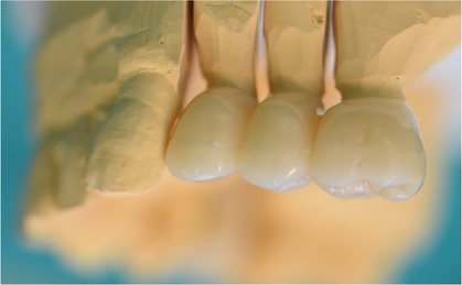
No impression copings or replicas are needed. All restorations were made of zirconia with an occlusal table made of composite.
The flexibility of the composite gives a more favourable stress absorption of the masticatory forces, see picture 5.
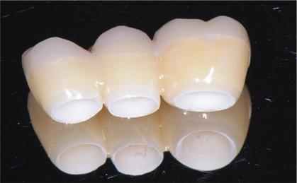
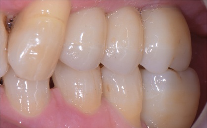
A bridge on three implants in regio 24-26 was made instead of three individual crowns to distribute the occlusal loads better. The antagonist to the implant in position 26 is also an implant (position 36). Since the 26 implant was only 4,5 x 9 mm it was decided to make the bridge restoration to distribute the load over the three implants, see pictures 6-9. A control X-ray was taken after final cementation, picture 10.
The implant in position 15 was a single tooth implant, see picture 11. In regio 36 another single tooth implant was placed, see picture 12. There is a small exposure of the implant but some creep of the soft tissue is expected over time. The control X-rays, pictures 13-14, show very stable marginal bone levels.
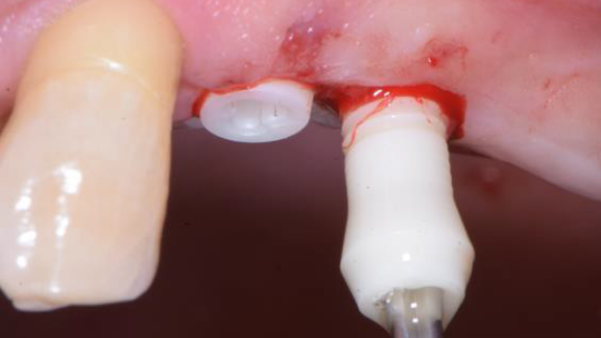
Picture 3:
Glass fiber posts cemented and prepared after 3 months of healing. Note the healthy soft tissues.
Picture 4:
The laboratory works on a plaster model just like crown & bridge. No impression copings or replicas are needed.
Picture 5:
The restoration is made of zirconia with the occlusal table made in composite to absorb the masticatory loads in a favourable way.
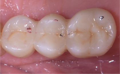
Picture 6:
Occlusion is checked.
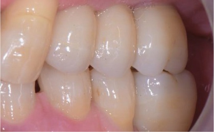
Picture 7:
Vestibular view.
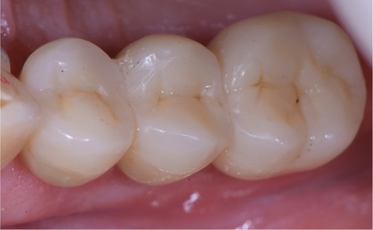
Picture 8:
Occlusal view.
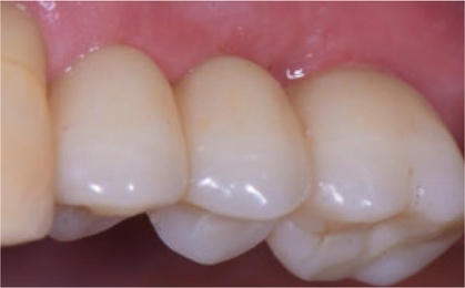
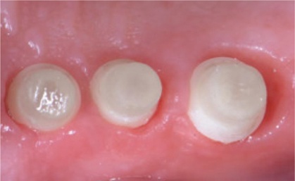
Picture 9:
Final result at the time of delivery.
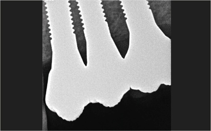
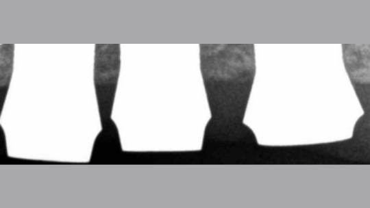
Picture 10:
Control X-ray after prosthetic delivery. Note the stable marginal bone levels.
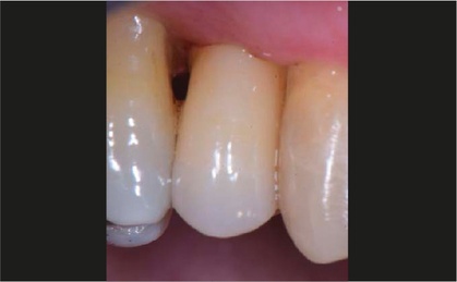
Picture 11:
Single tooth implant in position 15.
Picture 12:
Single tooth implant in position 36.
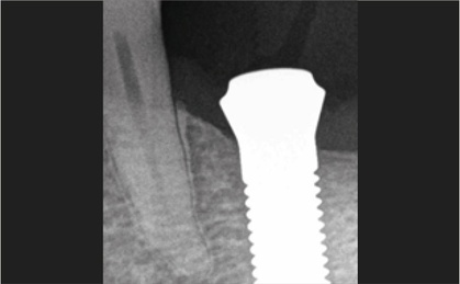
Picture 13:
Control X-ray at the time of implant placement and at the time of prosthetic delivery. Very stable marginal bone levels.
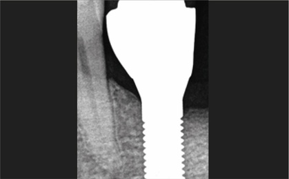
Picture 14:
Control X-ray at the time of implant placement and at the time of prosthetic delivery. Very stable marginal bone levels.
Conclusion
The Patent™ implant system offers a great prosthetic flexibility with the glass fiber post. Single units or bridge constructions can be made in a very efficient way using conventional dentistry techniques. The implant design allows total Bio-Integration with the rough surface in the bone and the machined surface on the transmucosal part. In combination with the design without any microgap deep down in the soft tissues, it gives very stable tissue levels, see X-rays in pictures 3, 10, 14, 14. The Patent™ Dental Implant System has shown survival rates on par with titanium and very favourable soft tissue response.
References
Roehling S, Schlegel K A, Woelfler H, Gahlert M. Performance and outcome of zirconia dental implants in clinical studies: a meta-analysis. Clin Oral Impl Res. 2018;29(Suppl. 16):135–153.
Brüll F, van Winkelhoff AJ, Cune MS. Zirconia dental implants: a clinical, radiographic, and microbiologic evaluation up to 3 years. Int J Oral Maxillofac Implants. 2014 Jul-Aug;29(4):914-20. Doi: 10.11607/jomi.3293
Becker J, John G, Becker K, Mainusch S, Diedrichs G, Schwarz F. Clinical performance of two- piece zirconium implants in the posterior mandible and maxilla: a prospective cohort study over 2 years. Clin. Oral Impl. Res. 28, 2017, 29–35 doi: 10.1111/clr.12610